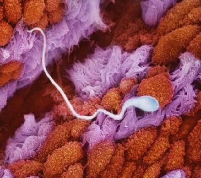
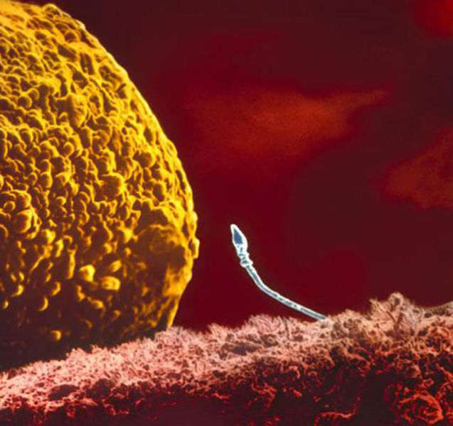
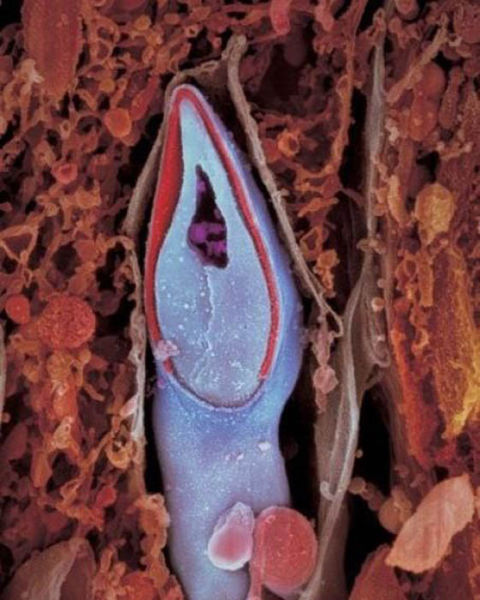
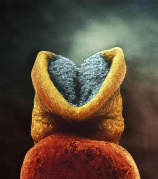
Swedesh photographer Lennart Nilsson spent 12 years of his life taking pictures of the foetus developing in the womb. These incredible photographs were taken with conventional cameras with macro lenses, an endoscope and scanning electron microscope. Nilsson used a magnification of hundreds of thousands and “worked” right in the womb. His first photo of the human foetus was taken in 1965.
1
5 weeks: Approximately 9 mm. You can now distinguish the face with holes for eyes, nostrils and mouth
40 days. Embryonic cells form the placenta. This organ connects the embryo to the uterine wall allowing nutrient uptake, waste elimination and gas exchange via the woman’s blood supply
The skeleton consists mainly of flexible cartridge. A network of blood vessels is visible through the thin skin






















3 comments:
wow boss u r great. how a chils is born
What a great work..... Hats off..
Thank you my Dil Se Desi Group I learn a lot of things here! I am thankful to my friend on Tagged Ron S for it was in one of his comments that I clicked this group! Be blessed always my friends!!!! Continue sharing knowledge!!
Post a Comment
Please Leave Your Precious Comments Here